Tel: (718) 648-8877 | Fax: (718) 648-4647
  • ABOUT US
    • Dr Sokolova
  • SERVICES
    • Arthritis Care
    • Joint Injections
    • Ultrasound
    • IV Therapy
    • PMS
    • Weight Loss
    • Wellness & rejuvenation
  • CONDITIONS WE TREAT
  • ANTI-AGING MEDICINE
    • Bioidentical Hormone Replacement
    • Next Generation Platelet Rich Plasma (PRP)
    • Weight Loss — Healthy Body
    • Anti-Inflammatory Diet: AID vs SAD
    • Fatigue Solutions
    • Leaky gut and autoimmunity
    • Metabolic Approach
    • Integrative Medicine
    • Genetic Consultation
  • PATIENT INFO
    • Office Hours & Location
    • Insurances Accepted
    • Policies
    • Patient Forms
  • ARTICLES

Ultrasound

  1. Home
  2. Medical Services
  3. Ultrasound

With ultrasound imaging of the musculoskeletal system?

Ultrasonography is a noninvasive medical test that helps physicians diagnose and treat the disease.

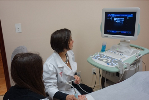
Ultrasound is safe and painless and produces pictures inside of the body using acoustic waves. Ultrasonography, also called ultrasound or sonography, involves the use of a small sensor (probe) and ultrasound gel is placed directly on the skin. High frequency sound waves transmitted from the probe into the body through the gel. The inverter collects the sounds that are reflected back, and then the computer uses the sound waves to create an image. Ultrasound studies do not use ionizing radiation (as used in x-rays), so no radiation exposure to the patient. Because ultrasound images are captured in real time, they can show the structure and motion of the internal organs of the body, and the blood flowing through the blood vessels.

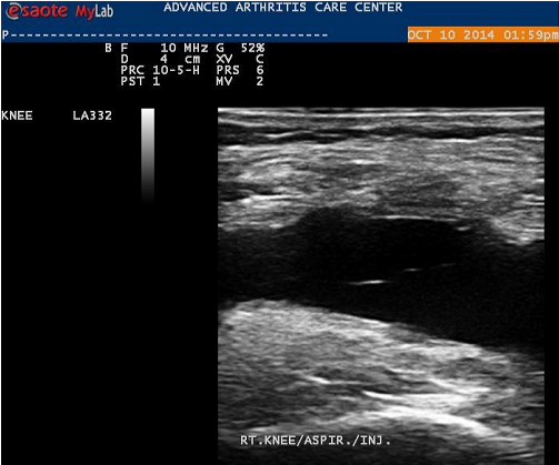
Ultrasound images of the musculoskeletal system, or Musculosceletal ultrasound, provide pictures of muscles, tendons, ligaments, joints and soft tissue throughout the body.

What are some common uses of the procedure?

With this technology, physicians can «see» inside the joint in real time and find indications of active inflammation or fluid-indicators that are often hard to find by palpation or visual assessment alone. There are also dynamic applications that allow physicians to assess a joint during movement. This is not possible using static modalities such as magnetic resonance imaging or computed tomography scans.

Ultrasound images are typically used to help diagnose:

  • tendon tears, or tendinitis of the rotator cuff in the shoulder, Achilles tendon in the ankle and other tendons throughout the body,
  • muscle tears, masses or fluid collections,
  • ligament sprains or tears,
  • inflammation or fluid (effusions) within the bursae and joints,
  • early changes, not seen on Xray, presence of erosions (joint damage) and degree of inflammation of rheumatoid and other types of arthritis. This is invaluable tool that allows us to tell different types of arthritis apart and determine how agressive arthritis is,
  • it allows us to see tiny depositions of crystals on cartlage and make a diagnosis of crystalline arthropathies, such as gout or pseudogout,
  • nerve entrapments such as carpal tunnel syndrome,
  • benign and malignant soft tissue tumors,
  • ganglion cysts,
  • hernias,
  • foreign bodies in the soft tissues (such as splinters or glass),
  • it is an exellent help in making sure that joint or other types of injections, such as carpal tunnel block, are done correctly. Guiaded joint injection is more effective because it ensures correct medication delivery. Besides, sono guiadence enables us to deliver medication into a deeply situated joints, such as hip, because without ultrasound guaidance this type of steroid injection has a high miss rate. Besides, even relatively simple injections, such as Supartz (viscous supplementation) injection into a knee, work better if joint fluid is removed before Supartz injection is done. Sono Guiadance makes it easy and simple and ensures the best possible result.

What will you experience during and after the procedure?

Ultrasound examinations are painless and easily tolerated by most patients. Musculoskeletal ultrasound examination is usually completed within 15 to 30 minutes but may occasionally take longer. When the examination is complete, you may be asked to dress and wait while the ultrasound images are reviewed. After an ultrasound examination, you should be able to resume your normal activities immediately.

Why may a follow-up exam be necessary?

Follow-up examinations may be necessary, and your doctor will explain the exact reason why another exam is requested. Sometimes a follow-up exam is done because a suspicious or questionable finding needs clarification with additional views or a special imaging technique. A follow-up examination may also be necessary so that any change in a known abnormality can be monitored over time. Follow-up examinations are sometimes the best way to see if treatment is working or if an abnormality is stable over time.

OUR GOAL

Our goal is simple yet, very complex at the same time – to deliver the best possible care in Integrative Rheumatology and Allergy that modern American medicine could offer, combining it with alternative holistic therapy.

MEDICAL SERVICES

  • Arthritis Care
  • Joint Injections
  • Ultrasound
  • PMS
  • Weight Loss
  • Intravenous Therapy
  • Wellness & rejuvenation
  • Solutions

CONTACT US

(718) 648-8877

For additional information about hours and operations or directions to our facility, please Contact Us at your convenience

Name *
Fill out this field
Email *
Please enter a valid email address.
Phone [optional]
Fill out this field
Massege *
Fill out this field
How much 19 + 13 = ?
Enter the equation result to proceed

This website uses cookies to improve your experience. If you continue to use this site, you agree with it. Policies